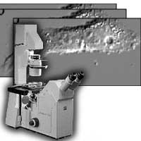
Differential interference contrast (DIC) microscopy, a method pioneered by Georges Nomarski, is widely used to study live biological specimens. However, to date, biologists only qualitatively interpret DIC microscope images. In this work, we describe a method to extract quantitative information from optically-sectioned DIC microscope images. Specifically, given a set of images of a specimen, we attempt to reconstruct the three-dimensional structure and refractive index distribution throughout the specimen.
The nonlinear nature of the DIC imaging process has hindered past attempts at quantitative analysis. Deconvolution of microscope images, also known as computational optical sectioning methods, is restricted to modalities, such as fluorescence. The image intensity, in such modalities, can be approximated as the convolution of a point spread function, or impulse response, with object source density, or irradiance. In contrast, the image seen in a DIC microscope is an interference image, and therefore the light amplitude has to be modelled, preserving phase information.
Our model, a generalized ray-tracer, uses energy conservation laws to compute the propagation of light through the object and the microscope. After calibrating the prism parameters, we use our model to estimate the specimen’s refractive index distribution. We trace rays, the normals to the surfaces of constant phase of the electric field, through inhomogeneous objects. We determine the intensity distribution at the image plane by computing the diffraction by the lens aperture, and the aberrations caused by the specimen’s self-occlusion. Therefore, we model multiple scatterings through the object, a better approximation than the first Born approximation of light scattered once by the object. Before using the model for the purpose of reconstruction, we validate its use by comparing real and simulated images of known objects.
We use an iterative non-linear optimization scheme to estimate the three-dimensional properties of the specimen. The specimen is represented by the refractive-index distribution across the volume enclosing it. We estimate discretely sampled points of this refractive-index distribution. Since the degrees of freedom of the system is large, we use a multi-resolution scheme to impose a regularization on the optimization. We represent the discrete refractive-index values with respect to a wavelet basis. At each iteration, we estimate more wavelet coefficients, and therefore estimate higher frequency components present in the specimen. To demonstrate that this method can estimate the refractive index distribution, we reconstruct a two-dimensional specimen.
current head
past staff
- Farhana Kagalwala
